1H and 19F on all systems + X nuclei for dual channel systems
Resolution:
Spinsolve 100 ULTRA
Linewidth at 50% <0.2 Hz
Linewidth at 0.55% <8 Hz
Linewidth at 0.11% <16 Hz
1H Sensitivity: >400:1 (Single channel for 1% Ethyl Benzene1)
Highest Sensitivity: >300:1 (Dual channel for 1% Ethyl Benzene1)
3D PFG gradients optimized for gradient-enhanced methods
Optional PFG gradients for diffusion spectroscopy (>0.5 T/m)
No cryogens
External Hardware Lock with no need for deuterated solvents
Unparalleled stability
Suitable for on-line reaction monitoring
Easy to operate
Available with automatic sample changer
Benchtop size and weight
Dimensions: 66 x 45 x 43 cm (25,9” x 17,7” x 16,9”)
Weight: 120 kg (265 lb)
1 Sensitivity measured in a single scan on a 1% ethylbenzene sample dissolved in deuterated chloroform. The signal used for the calculation is the one of the methylene group and the noise is calculated for a signal free region between the aromatic and the methylene signals.
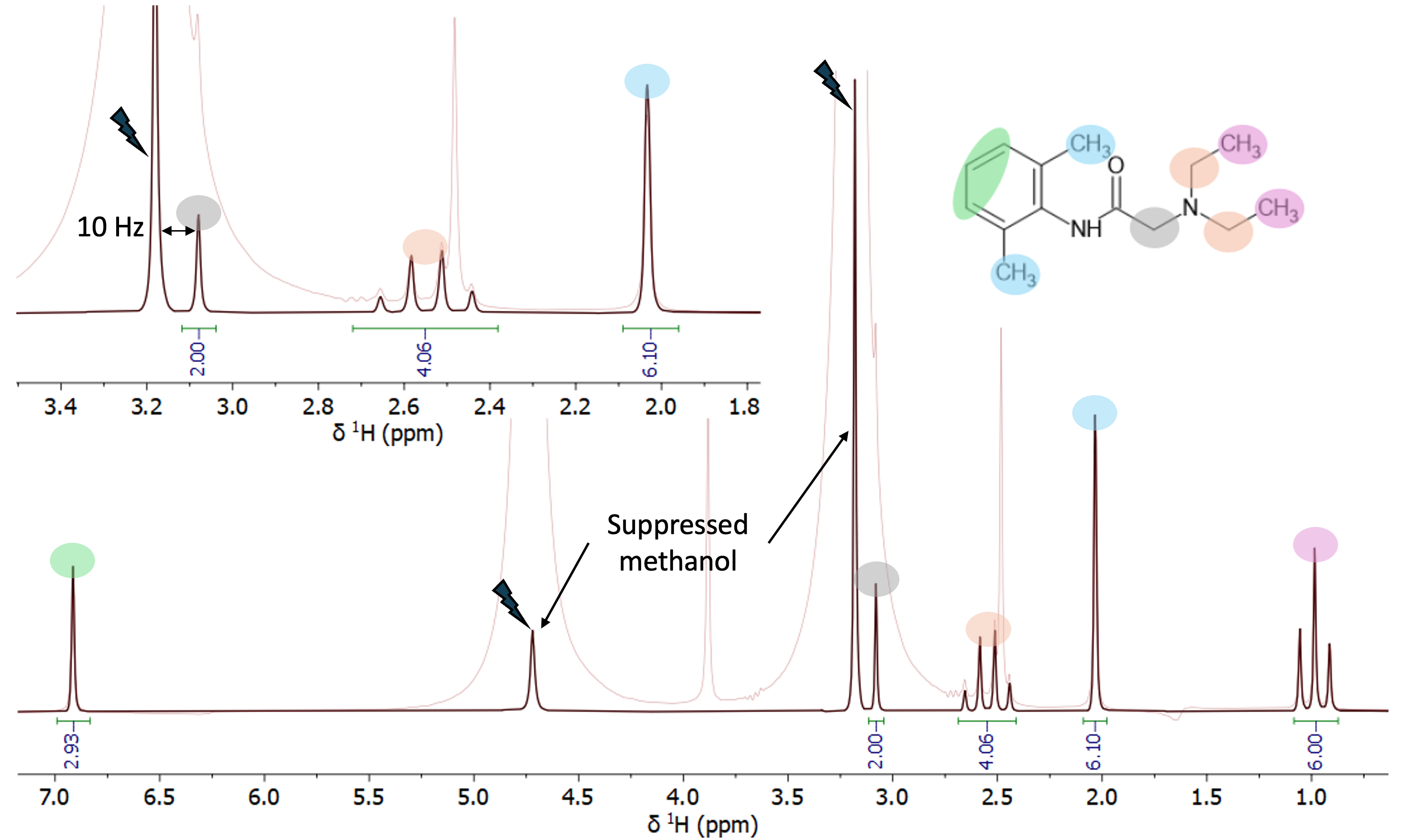
Discover the power of the solvent suppression methods
Measure your samples in regular protonated solvents with the accuracy obtained using deuterated solvents. By eliminating the sample work up required to exchange solvents, measurements can be done during your synthesis in just a few seconds. This is changing how NMR spectroscopy is being used in the chemistry lab for fast sample analysis.
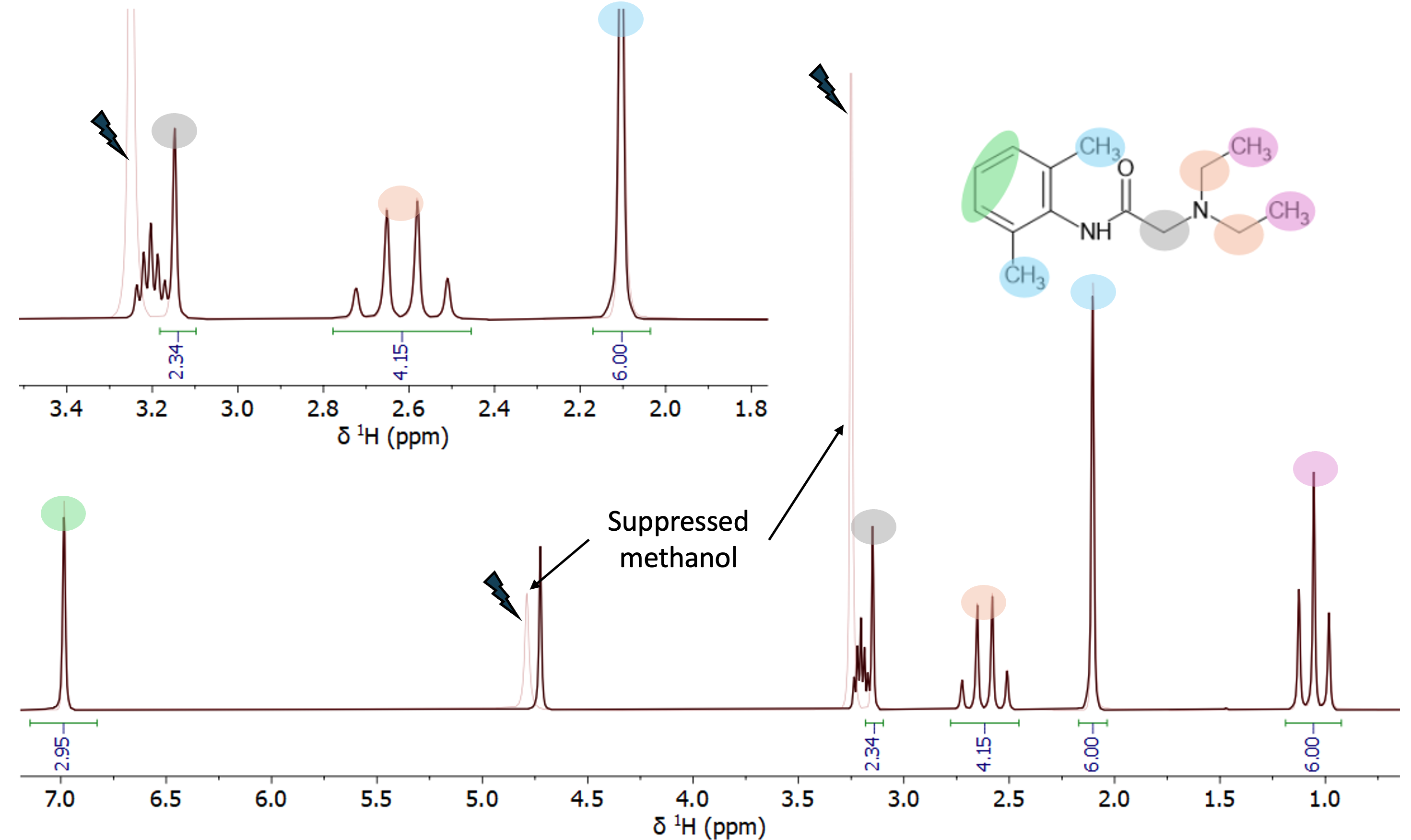
Spectra of Lidocaine (50 mMolar) in protonated methanol. The spectrum was acquired in one minute using a WET solvent suppression sequence targeting the two methanol signals. This highly selective suppression enables the lidocaine peak at 3.1 ppm—which fully overlaps with methanol in a standard 1D spectrum (light red)—to be baseline resolved and accurately integrated.
In the spectrum of lidocaine sample prepared in deuterated methanol, the intensity of the residual solvent signals is comparable to those of the residual signals of the protonated methanol in the solvent-suppressed spectrum (red trace). The inset shows the overlap of the multiplet from CH3 group of deuterated methanol and the lidocaine signal at 3.1 ppm. The solvent-suppressed spectrum enables more accurate integration of this lidocaine signal.
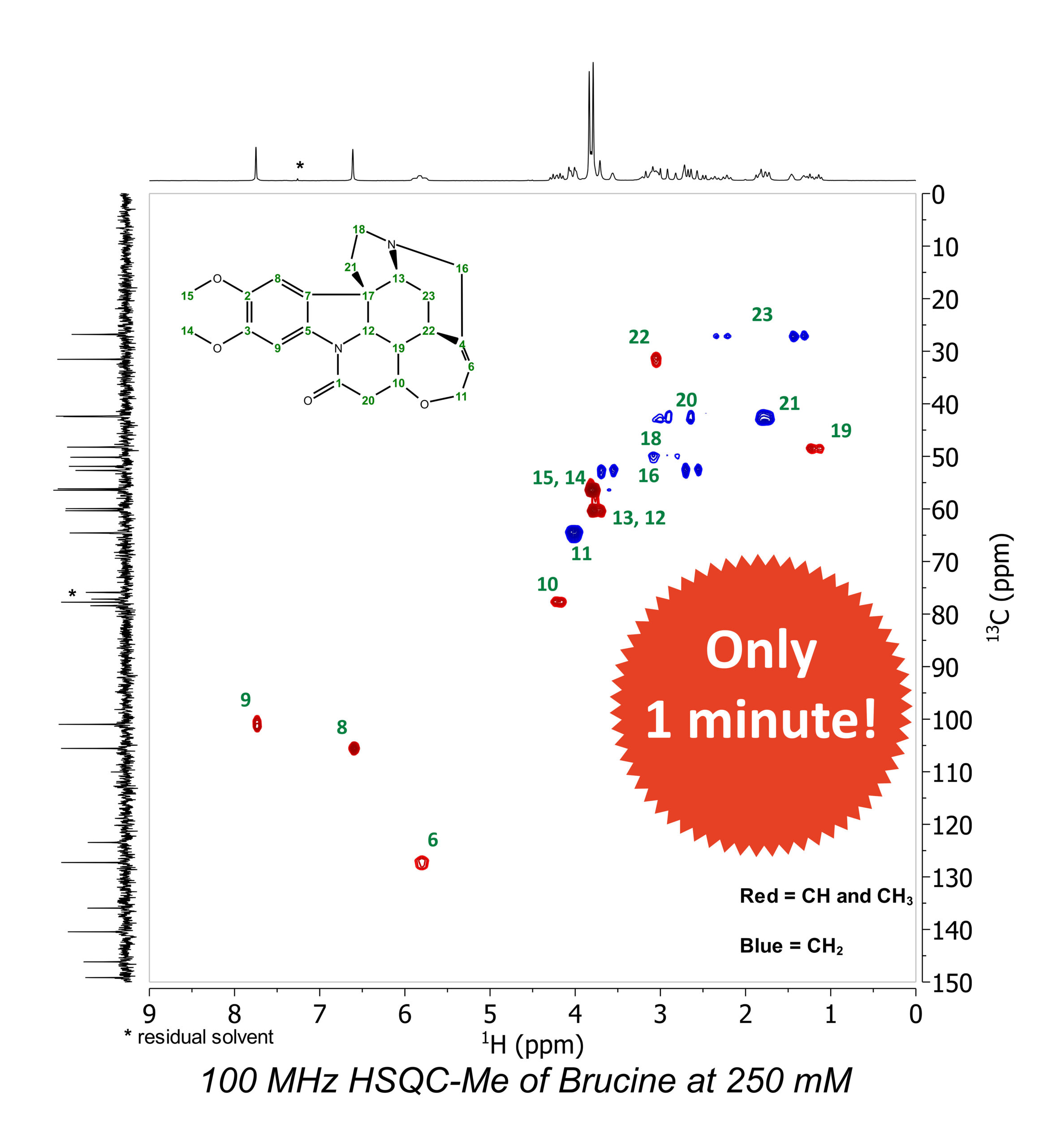
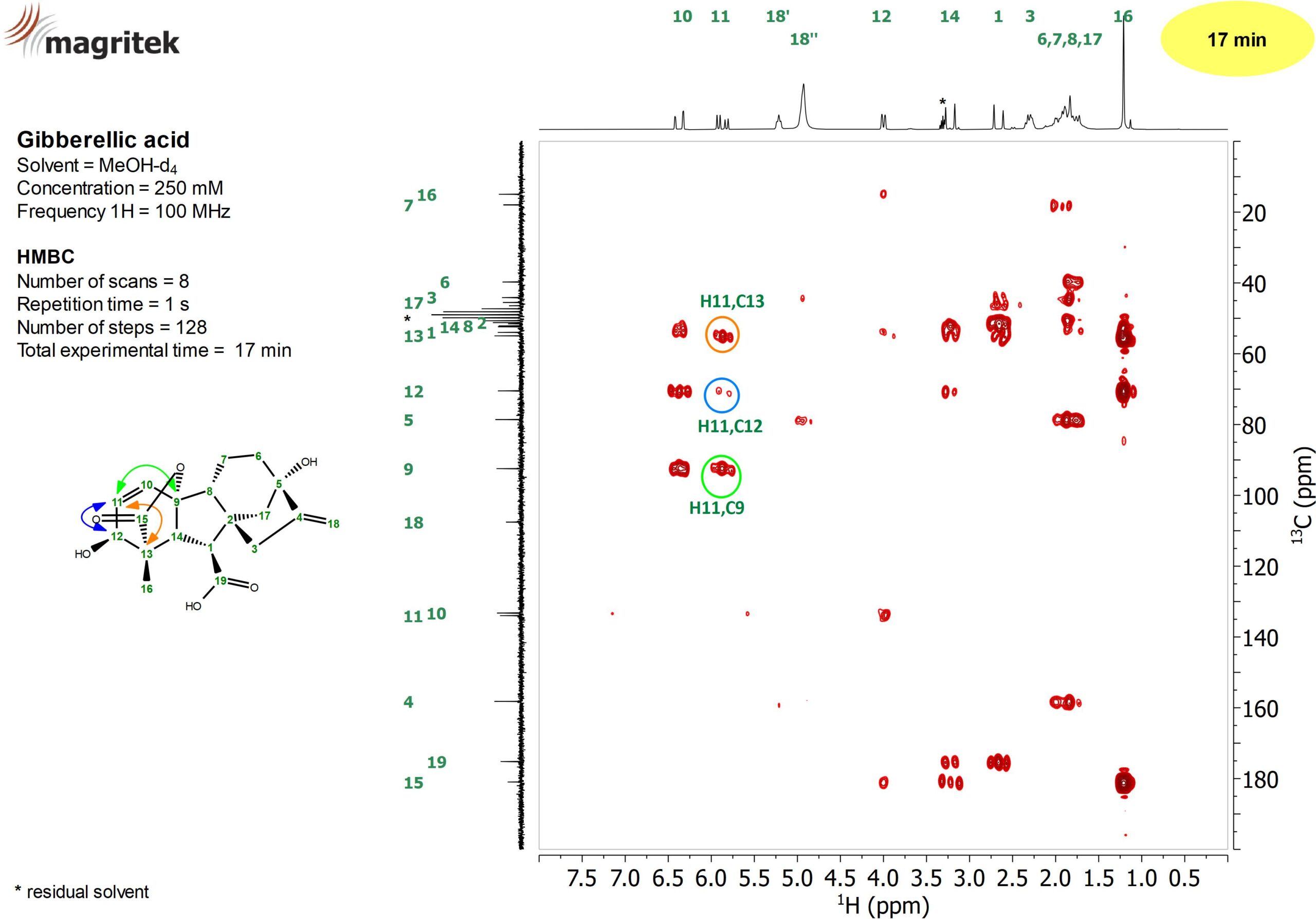
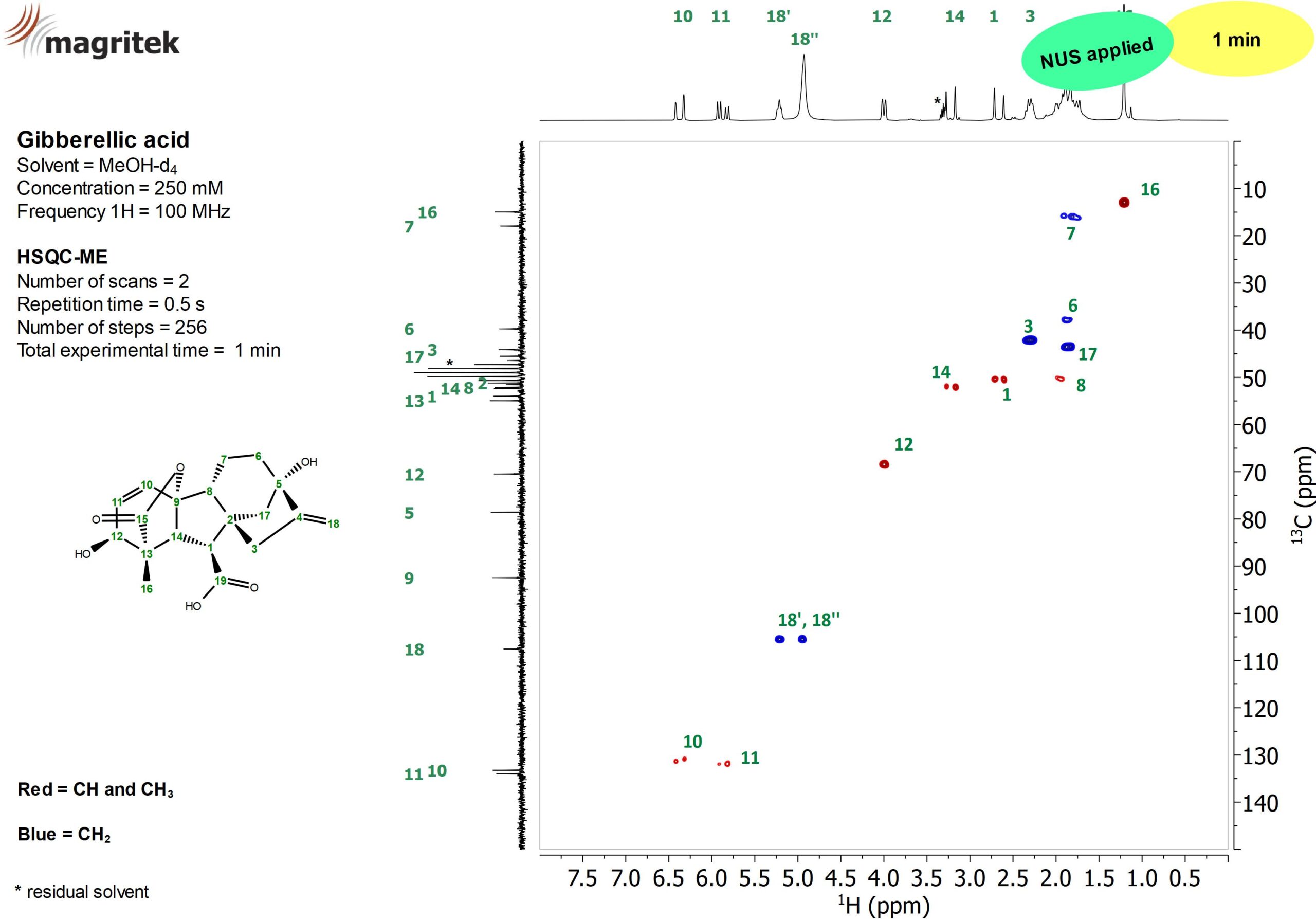
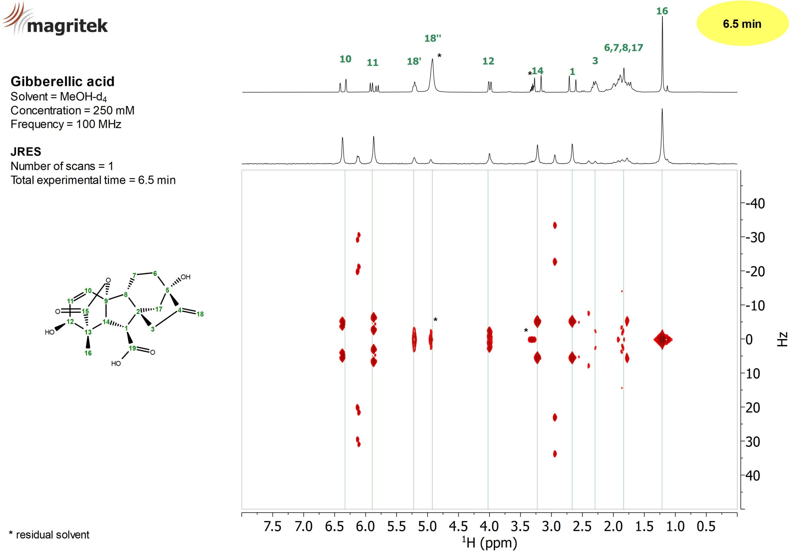
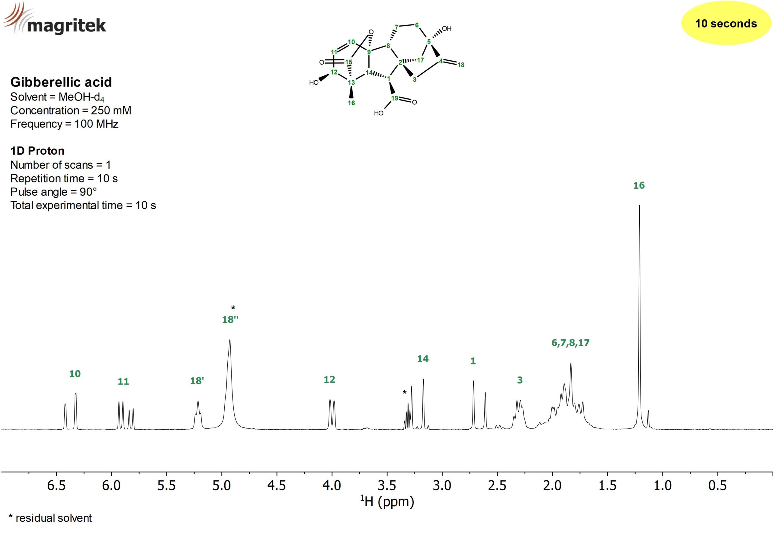
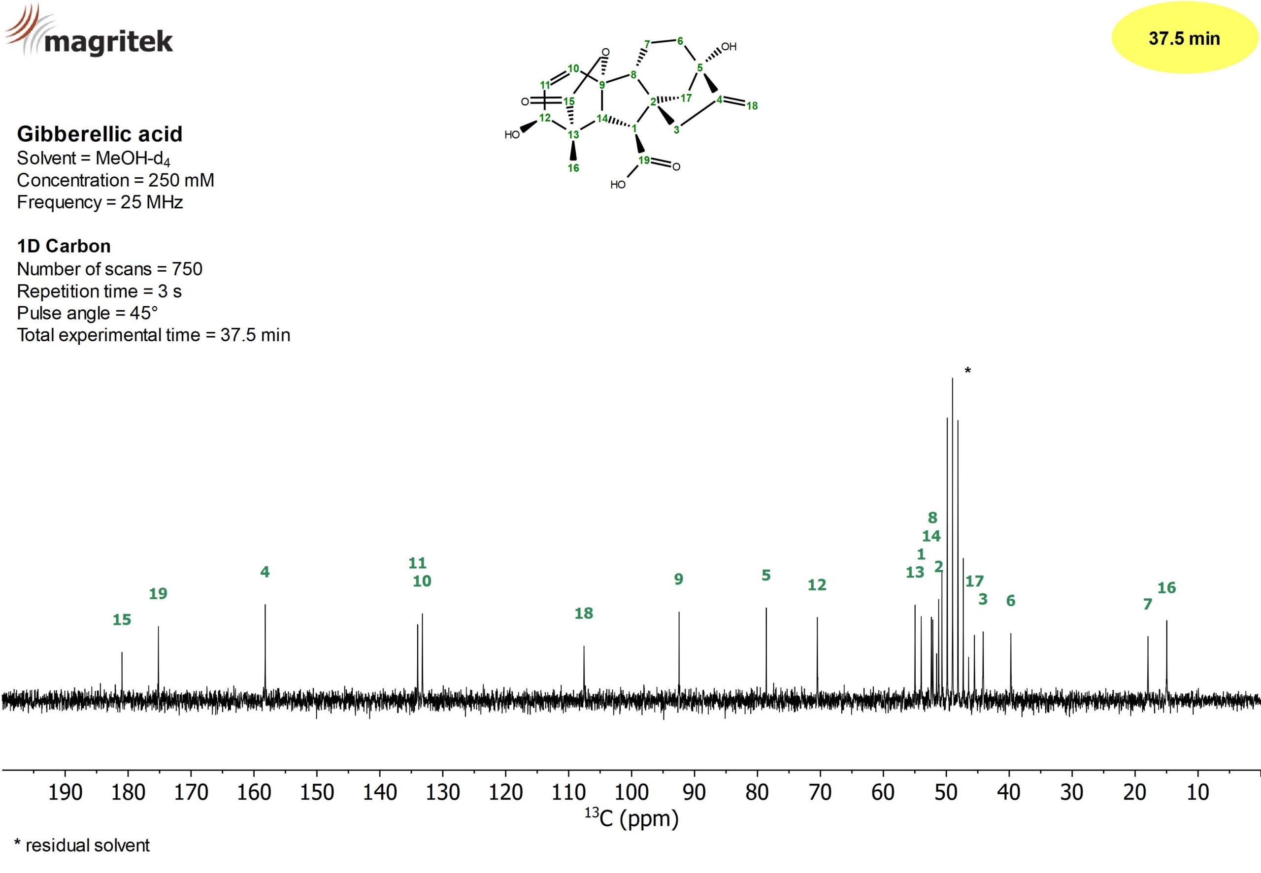
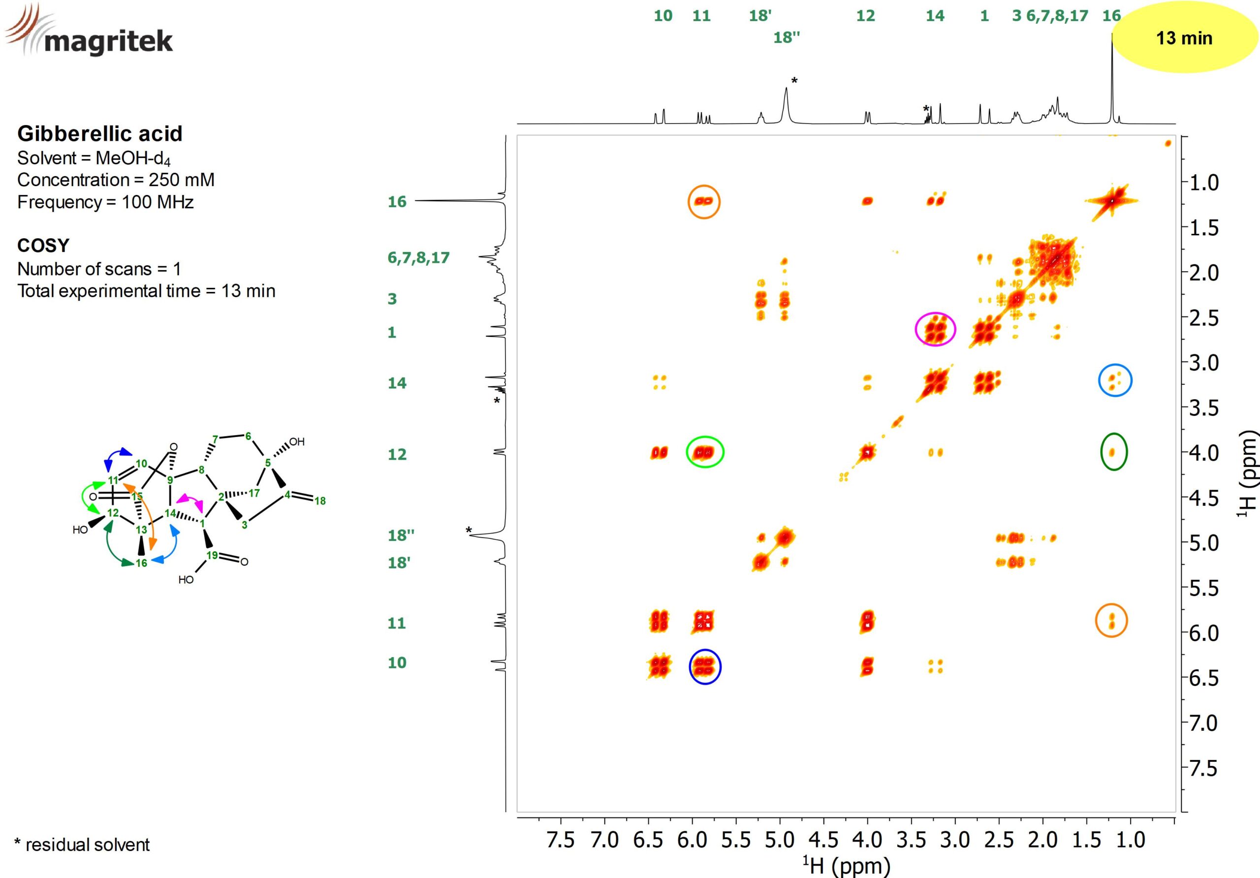
Fast and powerful, advanced multi-nuclear methods for structure confirmation
100 MHz NMR spectra of Gibberellic acid at 250 mMolar concentration
1H Sensitivity (*)(single channel): >400:1 for 1% Ethyl Benzene
1H Sensitivity (*)(dual channel): >300:1 for 1% Ethyl Benzene
Operating Temperature Range: 14° C to 28° C (57° F to 82° F)
(*) Sensitivity measured in a single scan on a 1% ethylbenzene sample dissolved in deuterated chloroform. The signal used for the calculation is the one of the methylene group and the noise is calculated for a signal free region between the aromatic and the methylene signals
Sample: Standard 5 mm OD NMR sample tubes, 7″ length
Minimum Sample Volume: 250 µl
External Hardware Lock with no need for deuterated solvents
3D PFG gradients optimized for gradient-enhanced methods
Optional PFG gradients for diffusion spectroscopy (>0.5 T/m)
Dimensions: 66 x 45 x 43 cm (25,9” x 17,7” x 16,9”)